
Imaging & Neuroscience
Radiology of posterior fossa syndrome
Group lead: Mina Langhein, MD · Co-lead: Stuart McAfee, PhD
What radiologists need to know about posterior fossa syndrome (PFS): the imaging signs of injury, how to assess risk before surgery, and what to look for during and after surgery.
What radiologists should know
Key points for radiologists
- PFS is a surgical complication in children having posterior fossa tumor resection.
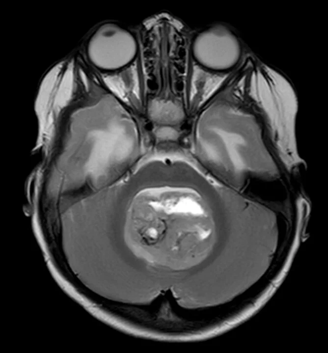
- Midline tumors, brainstem invasion, and superior cerebellar peduncle (SCP) invasion are the key radiological risk factors; medulloblastoma carries the highest risk.
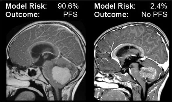
- Validated tools help assess the risk of PFS from preoperative MRI, but outcomes still depend on surgical technique.
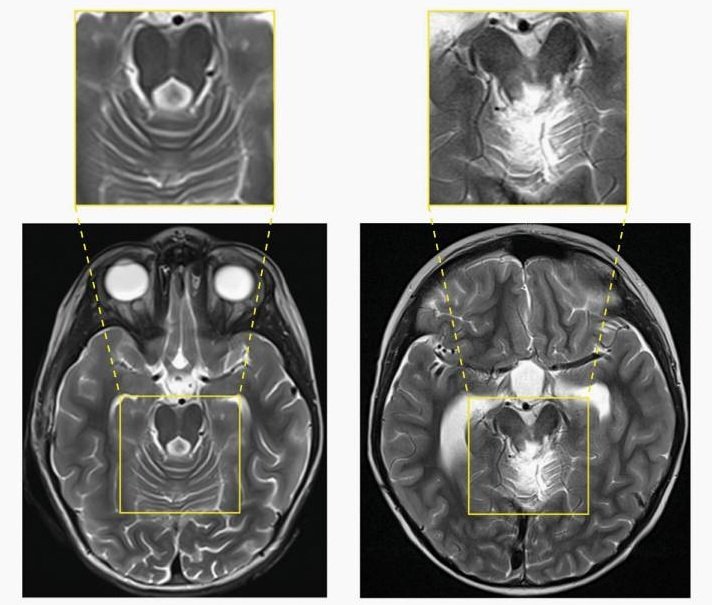
- Injury to structures around the fourth ventricle (dentate nuclei, SCPs, dorsal brainstem, and mesencephalic tegmentum) is an early postsurgical imaging feature associated with PFS.
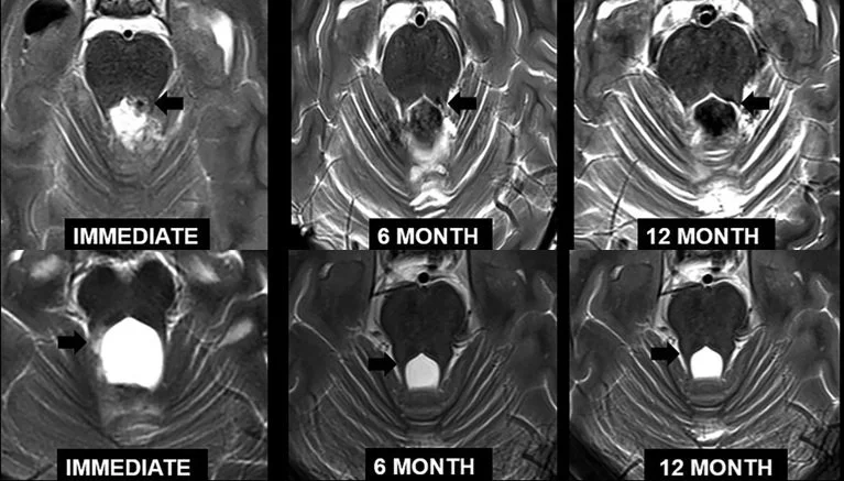
- Hypertrophic olivary degeneration is a well-recognized finding on surveillance MRI.
Bringing these findings to radiologists who rarely see the syndrome is a core aim of the group. Its members have prepared a review for the neuroradiology community, currently under peer review, summarizing where the evidence is strong and agreed.
Imaging correlates of injury
- The cerebellar outflow pathway. Postoperative T2 signal abnormality and diffusion restriction in the deep cerebellar nuclei, the SCPs, and their continuation in the posterior brainstem are more frequent in children who develop PFS.1, 2, 3
- The SCP as a bottleneck. All cerebellar outflow passes through the SCPs, so small lesions there can disrupt many functions at once.4 Bilateral diffusion abnormality of the dentato-thalamo-cortical tract (DTCT) is associated with complete mutism, and unilateral abnormality with milder speech impairment.1, 2, 5
- Midline, not hemispheric, injury. Tumors confined to the cerebellar hemispheres carry little risk of mutism, while midline tumors with surgical injury to the vermis or fastigial nuclei are more consistently associated with it.6, 7
- Symptom-specific findings. The side of DTCT injury on tractography predicts the side of limb dysmetria.5
For how these structures are thought to produce the syndrome, see our page on pathophysiology research.
Preoperative risk assessment
- Features. Brainstem invasion, midline tumor location, tumor size, involvement of the cerebellar efferent pathways, and medulloblastoma histology recur as risk factors across studies.8
- Models. The Liu model was the first based only on preoperative information, but its initial accuracy has not been replicated.9, 10 The Rotterdam model adds a sagittal measure of ventral brainstem displacement or invasion; in its development cohort (internal validation), accuracy was 87%, sensitivity 97%, and specificity 84%.11 The GOSH-Colorado-Stanford neural network model is available as a free online calculator.10 In a multicenter comparison, the Rotterdam model generalized better.12 Performance reported in a single cohort is usually higher than performance at new centers.
- Limits. The models depend on visual judgments of compression and invasion, for which agreement between radiologists varies.13 Radiomic and voxel-wise lesion-mapping models may avoid this but are not yet part of routine reporting.14, 15 Whatever the predicted risk, the Society recommends that resection be performed by an experienced pediatric neurosurgeon.16
Intraoperative MRI
Diffusion abnormality in the proximal efferent cerebellar pathway (dentate nucleus, SCP, and mesencephalic tegmentum) is the most accessible intraoperative marker of later PFS; bilateral involvement had a positive predictive value of 100% in one cohort, with weaker associations for the vermis and none for the middle cerebellar peduncle.2 Artifacts from gas, hemostatic material, and hematoma in the surgical cavity call for caution.17
Early postoperative MRI (24 to 72 hours)
Edema, gliosis, and diffusion changes in the vermis, dentate nuclei, and SCPs are well documented in children with PFS.1, 18 Perfusion imaging consistently shows frontal hypoperfusion,19, 20 and functional MRI shows altered cerebello-cerebral connectivity.21 Vascular imaging may be warranted in selected cases, given a reported case caused by basilar artery vasospasm.22
Follow-up imaging
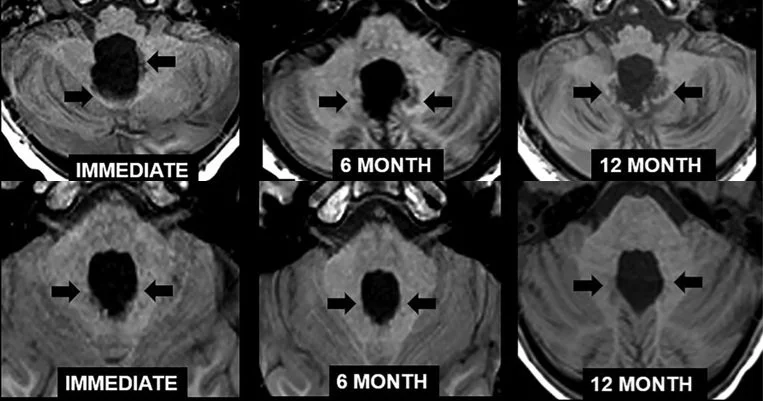
Diffusion tensor imaging shows reduced fractional anisotropy in the SCPs months after surgery in children with PFS.23, 24 Progressive bilateral T1 hyperintensity in the dentate nuclei, indicating dystrophic mineralization, is more common in children with PFS than in treatment-matched children without it.3 Hypertrophic olivary degeneration, reflecting trans-synaptic degeneration within the dentato-rubro-olivary pathway, is significantly associated with PFS, and bilateral involvement is highly specific.25, 26 In a longitudinal study, it was significantly more common in children with PFS, although only 40% of them developed it.3 Magnetization transfer imaging shows loss of tissue integrity downstream of the cerebellum, most marked in the ventral periaqueductal gray.27
Selected references
- Morris EB, Phillips NS, Laningham FH, et al. Proximal dentatothalamocortical tract involvement in posterior fossa syndrome. Brain. 2009;132(Pt 11):3087–3095. Link
- Avula S, Kumar R, Pizer B, et al. Diffusion abnormalities on intraoperative magnetic resonance imaging as an early predictor for the risk of posterior fossa syndrome. Neuro Oncol. 2015;17(4):614–622. Link
- Pinto SN, Scoggins MA, Patni T, et al. Longitudinal signal changes in the dentato-rubro-olivary pathway of patients with posterior fossa syndrome. Neuro-Oncol Pract. 2026. Link
- Albazron FM, Bruss J, Jones RM, et al. Pediatric postoperative cerebellar cognitive affective syndrome follows outflow pathway lesions. Neurology. 2019;93(16):e1561–e1571. Link
- Ji Q, McAfee SS, Scoggins M, et al. Cerebellar mutism syndrome and dentato-thalamo-cortical tract disruption in diffusion tractography following surgery for medulloblastoma. Radiology. 2024;311(2):e232521. Link
- Khan RB, Patay Z, Klimo P, et al. Clinical features, neurologic recovery, and risk factors of postoperative posterior fossa syndrome and delayed recovery: a prospective study. Neuro Oncol. 2021;23(9):1586–1596. Link
- Grønbæk JK, Wibroe M, Toescu S, et al. Postoperative speech impairment and surgical approach to posterior fossa tumours in children: a prospective European multicentre cohort study. Lancet Child Adolesc Health. 2021;5(11):814–824. Link
- Obdeijn IV, van Baarsen KM, Avula S, et al. Neuroimaging of postoperative pediatric cerebellar mutism syndrome: a systematic review. Neurooncol Adv. 2025;7(1):vdae212. Link
- Liu JF, Dineen RA, Avula S, et al. Development of a pre-operative scoring system for predicting risk of post-operative paediatric cerebellar mutism syndrome. Br J Neurosurg. 2018;32(1):18–27. Link
- Sidpra J, Marcus AP, Löbel U, et al. Improved prediction of postoperative pediatric cerebellar mutism syndrome using an artificial neural network. Neurooncol Adv. 2022;4(1):vdac003. Link
- Dhaenens BAE, van Veelen MLC, Catsman-Berrevoets CE. Preoperative prediction of postoperative cerebellar mutism syndrome: validation of existing MRI models and proposal of the new Rotterdam pCMS prediction model. Childs Nerv Syst. 2020;36(7):1471–1480. Link
- Sidpra J, Toescu SM, Yamaki V, et al. Artificial intelligence improves the preoperative prediction of cerebellar mutism syndrome: a multinational, multi-reader study and practice recommendations from the Posterior Fossa Society [abstract]. Neuro-Oncology Pediatrics. 2025;1(Suppl 1):wuaf001.163. Link
- Laustsen AF, Dineen R, Ilginiene J, et al. Interobserver variability in assessing preoperative imaging biomarkers for cerebellar mutism syndrome: a multiobserver pilot study. Pediatr Radiol. 2025;55(9):1915–1926. Link
- Yang W, Yang P, Li Y, et al. Presurgical MRI-based radiomics models for predicting cerebellar mutism syndrome in children with posterior fossa tumors. J Magn Reson Imaging. 2023;58(6):1966–1976. Link
- Yang W, Li Y, Ying Z, et al. A presurgical voxel-wise predictive model for cerebellar mutism syndrome in children with posterior fossa tumors. NeuroImage Clin. 2023;37:103291. Link
- Toescu SM, Pizer B, Gump W, et al. Toward reducing the risk of cerebellar mutism syndrome: consensus statement from the Posterior Fossa Society. J Neurosurg Pediatr. 2025;36(6):789–797. Link
- Avula S, Jaspan T, Pizer B, et al. Comparison of intraoperative and post-operative 3-T MRI performed at 24–72 h following brain tumour resection in children. Neuroradiology. 2021;63(8):1367–1376. Link
- Skye J, Bruss J, Toescu S, et al. Neuroanatomy of cerebellar mutism syndrome: the role of lesion location. Brain Commun. 2024;6(4):fcae197. Link
- Miller NG, Reddick WE, Kocak M, et al. Cerebellocerebral diaschisis is the likely mechanism of postsurgical posterior fossa syndrome in pediatric patients with midline cerebellar tumors. AJNR Am J Neuroradiol. 2010;31(2):288–294. Link
- Ahmadian N, van Baarsen KM, Robe P, Hoving EW. Association between cerebral perfusion and paediatric postoperative cerebellar mutism syndrome after posterior fossa surgery: a systematic review. Childs Nerv Syst. 2021;37(9):2743–2751. Link
- McAfee SS, Robinson G, Gajjar A, et al. Secondary cerebro-cerebellar and intra-cerebellar dysfunction in cerebellar mutism syndrome. Neuro Oncol. 2024;26(9):1700–1711. Link
- Deghedy M, Pizer B, Kumar R, et al. Basilar artery vasospasm as a cause of post-operative cerebellar mutism syndrome. Case Rep Pediatr. 2022;2022:9148100. Link
- Toescu SM, Bruckert L, Jabarkheel R, et al. Spatiotemporal changes in along-tract profilometry of cerebellar peduncles in cerebellar mutism syndrome. NeuroImage Clin. 2022;35:103000. Link
- Vedantam A, Stormes KM, Gadgil N, et al. Association between postoperative DTI metrics and neurological deficits after posterior fossa tumor resection in children. J Neurosurg Pediatr. 2019;24(4):364–370. Link
- Patay Z, Enterkin J, Harreld JH, et al. MR imaging evaluation of inferior olivary nuclei: comparison of postoperative subjects with and without posterior fossa syndrome. AJNR Am J Neuroradiol. 2014;35(4):797–802. Link
- Avula S, Spiteri M, Kumar R, et al. Post-operative pediatric cerebellar mutism syndrome and its association with hypertrophic olivary degeneration. Quant Imaging Med Surg. 2016;6(5):535–544. Link
- McAfee SS, Zhang S, Zou P, et al. Fastigial nuclei surgical damage and focal midbrain disruption implicate PAG survival circuits in cerebellar mutism syndrome. Neuro Oncol. 2023;25(2):375–385. Link
Get involved
Society members can join this group by contacting the Imaging & Neuroscience lead or co-lead or writing to info@posteriorfossasociety.org. Not yet a member? Membership is open to clinicians and researchers working on posterior fossa syndrome.